


A thin uterine lining can matter for IVF success, but it is not always a barrier to pregnancy. The first priority is finding treatable causes, such as adhesions or infection, rather than assuming thickness alone decides the outcome. If your lining stays thin despite adequate estrogen, ask your clinic about hysteroscopy, the diagnostic gold standard for structural causes, before considering adjuvant therapies.
TL;DR:
- A thin endometrial lining is often caused by structural issues like adhesions or infections, which require specific treatment before considering adjuvants.
- Confirm the timing of ultrasound measurement and perform hysteroscopy if the lining remains thin despite normal estrogen exposure, as it is the gold standard diagnosis.
- Treatments such as hysteroscopic adhesiolysis and addressing infections can improve lining quality, potentially reducing the need for experimental therapies like PRP or G-CSF.
- Adjuvants like PRP show a modest increase in lining thickness but have limited evidence linking them directly to higher live birth rates.
- Lifestyle factors such as smoking cessation and maintaining good uterine blood flow support lining health but do not replace targeted structural or inflammatory treatments.
Table of Contents
- What is a thin uterine lining and how is it measured
- Causes of a thin endometrium: hormonal versus structural
- A practical diagnostic pathway: which tests come first
- Stepwise treatment options and what the evidence actually shows
- What the best studies and reviews say about outcomes
- Decision checklist and questions for your clinician
- Impact of thin uterine lining on natural conception versus IVF
- Lifestyle and natural cycle factors that influence lining thickness
- Endometrial receptivity testing and biomarkers in thin lining cases
- Regenerative therapies beyond PRP: where the research stands
- Prognosis: what to expect based on severity and response
- Supporting patients through diagnosis and treatment
- A clinician’s perspective on avoiding futile cycles
- How Barbados Fertility Centre supports refractory thin lining cases
- Key studies, reviews and guidelines used for this article
- Sources
- FAQ
What is a thin uterine lining and how is it measured
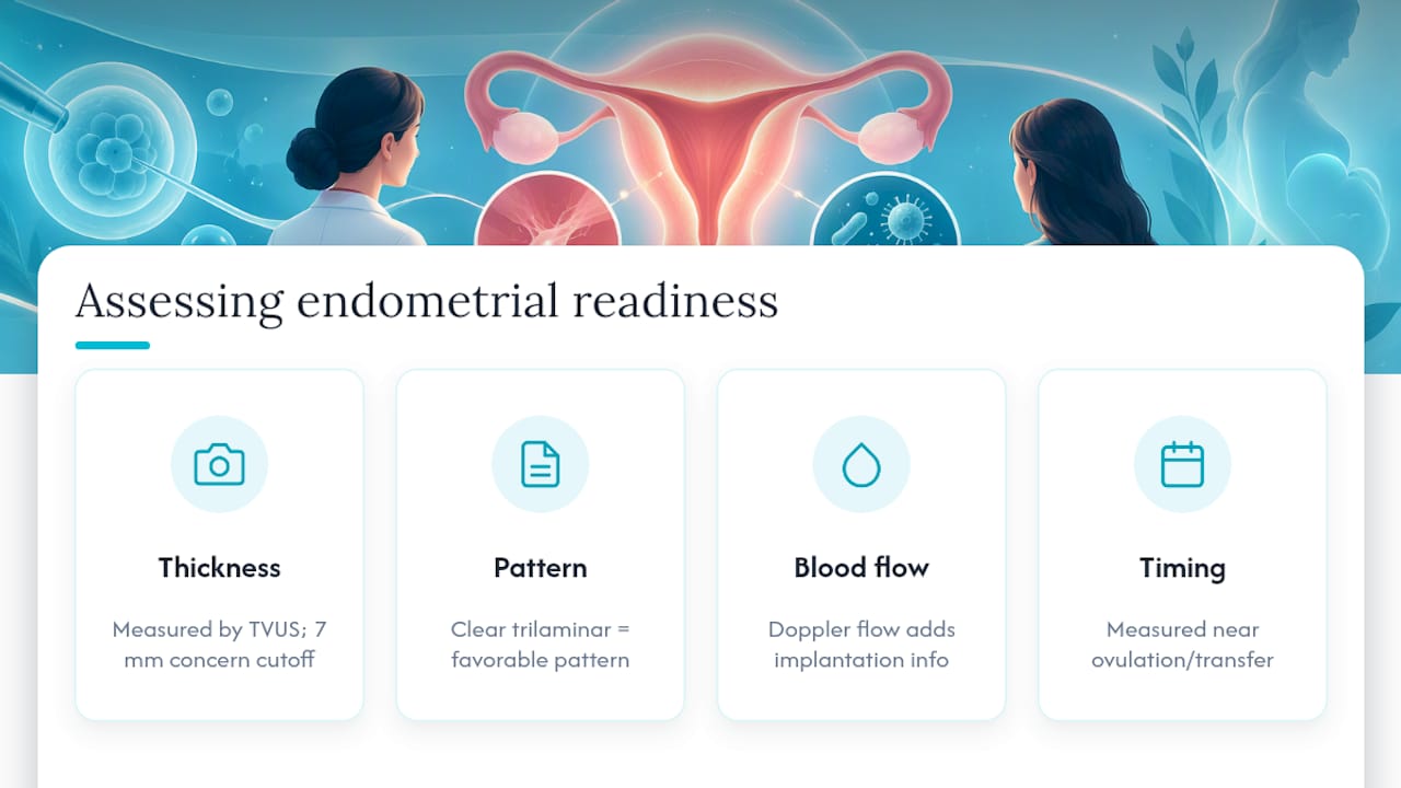
Endometrial thickness, often shortened to EMT, is measured with transvaginal ultrasound (TVUS), usually just before ovulation or before an embryo transfer. Clinicians look at the lining from front wall to back wall on a mid-sagittal image, and the reading depends heavily on timing within the cycle.
Most studies and protocols use a 7 mm cutoff as the point where clinicians start to worry, though the exact threshold varies by clinic and by whether the cycle is natural or hormonally programmed. A lining measured too early or too late in the cycle can look thinner than it truly is at the time that matters for implantation.
Thickness alone is a surrogate marker. Pattern, that is, whether the lining shows a clear trilaminar appearance, and blood flow to the area also carry information that a single number cannot capture.
Causes of a thin endometrium: hormonal versus structural
Not every thin lining has the same origin, and the cause determines the fix. Some cases trace back to a scarred cavity that estrogen cannot expand, while others reflect a hormonal or vascular problem that responds to different treatment altogether.
- Structural causes: intrauterine adhesions (Asherman syndrome), prior dilation and curettage, uterine surgery, radiation therapy, or polyps and fibroids that distort the cavity.
- Functional and hormonal contributors: a poor estrogen response, effects from clomiphene citrate, age-related thinning, and impaired uterine blood flow or angiogenesis.
- Inflammatory causes: chronic endometritis, a low-grade infection of the lining that is often missed and is treatable with antibiotics.
Poor subendometrial perfusion and weak blood vessel formation are recognized as key contributors to persistently thin linings that do not respond well to estrogen alone. That distinction matters when choosing between hormone adjustments and procedures aimed at restoring the cavity or its blood supply.
A practical diagnostic pathway: which tests come first
Working through a thin lining methodically saves time and avoids repeating cycles that were never going to succeed. A reasonable sequence looks like this:
- Confirm timing first. Repeat the transvaginal ultrasound at the correct point in the cycle before assuming the lining is truly thin.
- Add sonohysterography (SHG) if the cavity outline looks irregular, since saline infusion during ultrasound reveals polyps, adhesions, or septa that a standard scan can miss.
- Refer for hysteroscopy when the lining stays thin despite adequate estrogen exposure. Hysteroscopy is the gold standard for diagnosing intrauterine adhesions, and it allows the surgeon to treat them in the same procedure.
- Consider an endometrial biopsy when chronic endometritis is suspected, since a short course of targeted antibiotics can resolve it.
- Use 3D ultrasound, MRI, or Doppler studies in more complex or refractory cases where blood flow to the lining is in question.
Skipping straight to medications without ruling out a scarred or infected cavity is a common reason treatments seem to fail.
Stepwise treatment options and what the evidence actually shows
When a structural cause like adhesions is found, treating it directly, through hysteroscopic adhesiolysis followed by estrogen support, addresses a clear mechanical problem rather than masking it. This step generally comes before trying adjuvant therapies.
For cases without a clear structural cause, several options exist, though their evidence varies:
- Estrogen supplementation: oral, vaginal, or transdermal estrogen is the first-line approach to build the lining in a programmed cycle, with dose adjusted to response.
- Platelet-rich plasma (PRP): an intrauterine infusion of the patient’s own concentrated platelets, used when standard estrogen protocols fail.
- Granulocyte colony-stimulating factor (G-CSF): infused into the uterine cavity to try to stimulate lining growth.
- Sildenafil: used vaginally in some protocols to improve blood flow to the uterus.
- Aspirin and growth hormone: added in some regimens, though supporting data is inconsistent.
A meta-analysis of PRP trials found a pooled increase in endometrial thickness of roughly 0.9 to 1.3 millimeters, with a marginal improvement in clinical pregnancy rates and only limited data on live birth. That gap between a thicker lining and an actual birth is the central caveat with nearly every adjuvant on this list.
PRP requires drawing and processing the patient’s own blood before infusion, adding a procedure and processing time to the cycle. G-CSF and sildenafil are generally well tolerated, but none of these treatments are risk-free or guaranteed, and they add cost and time to a treatment plan that a reversible structural fix might solve outright.
What the best studies and reviews say about outcomes
Large multicenter studies complicate the simple idea that a thicker lining always means a better outcome. Practices differ by center: some routinely cancel or delay transfer when the lining is thin, while others proceed anyway, and that difference in practice affects which patients end up in the data at all. A Yale-reported multicenter study noted that centers proceeding with transfer despite a lining under 7 mm did not necessarily see worse outcomes, suggesting the underlying cause of the thin lining, not the number itself, often drives the result.
Meta-analyses of PRP and related interventions show a real, if modest, increase in measured thickness, alongside a marginal improvement in clinical pregnancy rates. Live birth, the outcome patients actually care about, remains far less certain across these trials. Honest counseling means separating what moves a millimeter on an ultrasound screen from what actually changes the odds of taking home a baby.
Decision checklist and questions for your clinician
Before agreeing to any adjuvant therapy, work through a short checklist with your care team.
- Confirm ultrasound timing was correct before accepting a thin-lining diagnosis.
- Ask for sonohysterography if cavity anatomy has not been directly visualized.
- Request hysteroscopy and biopsy to rule out adhesions or chronic endometritis before adding PRP or G-CSF.
- Ask what percentage improvement in thickness, and in live birth, is realistic for any therapy suggested.
Most structural issues can be addressed within one to two cycles of diagnosis and treatment; if the lining stays thin after that, a second opinion at a clinic with full diagnostic capability is reasonable.
Pro Tip: Ask your clinician to separate expected gain in millimeters from expected change in pregnancy odds before agreeing to any adjuvant treatment.
Impact of thin uterine lining on natural conception versus IVF
Thin lining affects natural conception and assisted reproduction differently, largely because of how each is monitored. In a natural cycle, thickness is rarely checked unless a woman is already being investigated for infertility, so many cases of thin lining outside a fertility clinic go unnoticed and unconfirmed.
In IVF and frozen embryo transfer cycles, the lining is measured deliberately, often repeatedly, before the embryo transfer date is set. This creates more opportunity to catch and address a thin lining, but it also means IVF patients face a clinical decision point that natural conception simply does not surface: proceed with transfer, delay, or cancel the cycle.
Programmed frozen cycles, where estrogen is given synthetically, appear to be more sensitive to thin lining effects than natural or modified natural cycles, based on the same multicenter data discussed earlier. That difference is one reason some clinics switch protocols, moving a patient from a programmed cycle to a natural or modified natural one, before trying medication-based adjuvants. The practical takeaway is that the setting in which thickness is measured, and how the cycle itself is managed, shapes the clinical response as much as the number on the screen.
Lifestyle and natural cycle factors that influence lining thickness
Endometrial thickness responds to more than hormones and procedures. Blood flow to the uterus, which underpins lining growth, is sensitive to several everyday factors.
Smoking is consistently associated with reduced uterine blood flow, and stopping is one of the few lifestyle changes with a clear physiological rationale for supporting a healthier lining. Chronic stress can disrupt the hormonal signaling that drives the menstrual cycle, though its direct effect on lining thickness specifically is less well defined than its broader effect on cycle regularity.
Moderate exercise supports healthy circulation generally, but very intense or excessive training can suppress reproductive hormone levels in some women, which may work against lining growth rather than for it. Nutrition plays a supporting role: adequate iron, protein, and overall caloric intake matter for tissue growth generally, though no specific diet has been shown to reliably thicken the endometrium on its own.
None of these factors replace a medical workup when the lining stays thin cycle after cycle. They matter most as background support alongside, not instead of, addressing a structural or inflammatory cause.
Endometrial receptivity testing and biomarkers in thin lining cases
Endometrial receptivity assays are tests designed to assess whether the lining is in the right hormonal state to accept an embryo, independent of its thickness. These tests typically involve a biopsy timed to the expected transfer window, analyzing gene expression patterns associated with receptivity.
For patients with a thin lining, receptivity testing addresses a different question than thickness measurement does. A lining can be thin and still receptive, or of adequate thickness and poorly receptive, which is part of why thickness alone is an incomplete predictor of transfer success. Biomarker approaches remain an area of active investigation rather than a routine, universally recommended step, and their role is generally considered alongside, not as a replacement for, structural and inflammatory workup.
Clinicians tend to reserve receptivity testing for patients who have already had structural causes ruled out and have experienced repeated implantation failure despite a technically adequate lining. It is one more tool in a broader diagnostic pathway rather than a stand-alone answer to a thin lining.
Regenerative therapies beyond PRP: where the research stands
Beyond platelet-rich plasma and growth hormone, researchers are exploring stem cell based approaches to regenerate a thin or damaged endometrium, particularly in cases involving significant scarring from Asherman syndrome. These approaches typically involve isolating stem cells, often from bone marrow or menstrual blood, and infusing them into the uterine cavity with the goal of stimulating tissue repair and new blood vessel growth.
This work remains in early research stages. Current evidence comes largely from small studies and case series rather than the large randomized trials available for PRP, and stem cell protocols vary considerably between research centers in terms of cell source, preparation, and delivery method. Patients considering these approaches should understand they fall outside standard, widely validated care and are best pursued, if at all, within a formal research or specialized program rather than as a routine clinical offering.
The underlying rationale, targeting the vascular and tissue regeneration deficits seen in refractory thin linings, is biologically reasonable given what is known about impaired perfusion in these cases. Reasonable rationale is not the same as proven benefit, and patients should be counseled accordingly before pursuing these still-experimental routes.
Prognosis: what to expect based on severity and response
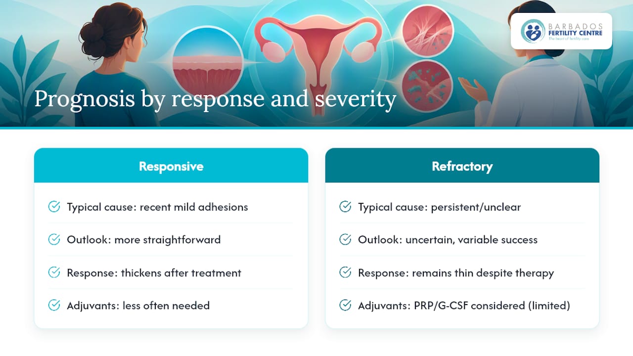
Outcomes for thin uterine lining vary considerably depending on the underlying cause and how the lining responds to initial treatment. A thin lining caused by mild, recently formed adhesions that resolve fully after hysteroscopic treatment carries a different outlook than a lining that stays thin despite adequate estrogen and a clear cavity on imaging.
Patients whose lining thickens promptly after treating a structural or inflammatory cause tend to have a more straightforward path forward, since the underlying problem has been directly addressed rather than worked around. Refractory cases, where the lining remains thin despite hysteroscopic correction and estrogen optimization, carry more uncertainty, and this is where the evidence for adjuvants like PRP or G-CSF becomes most relevant, even though that evidence is still developing.
Severity matters too: a lining that reaches 6.5 mm after treatment sits in a different risk category than one that plateaus at 4 mm despite every reasonable intervention. Realistic counseling means acknowledging that some cases will need to proceed with transfer at a less than ideal thickness, some will benefit from switching cycle type, and a smaller number will need to consider alternate paths such as gestational surrogacy if the lining truly cannot be improved. None of these outcomes are failures of effort; they reflect the biological reality that not every case of thin lining has a full fix available yet.
Supporting patients through diagnosis and treatment
A thin lining diagnosis often arrives at an already stressful point in fertility treatment, and it can feel like one more obstacle in a process that already demands a great deal of patience. Clear, honest communication about what is known and unknown helps patients make decisions without false reassurance or unnecessary alarm.
Framing the diagnosis around what is treatable, rather than around the number itself, tends to reduce distress. A patient told that hysteroscopy found and corrected an adhesion is in a fundamentally different emotional position than one told only that their lining is thin with no clear plan.
Clinics that build in time for questions, explain the reasoning behind each recommended test, and set realistic expectations about adjuvant therapies tend to support better decision-making and less anxiety around each cycle. Encouraging patients to ask directly about expected thickness gains versus expected changes in pregnancy odds, as covered earlier, also gives them a concrete way to participate in decisions rather than simply following instructions.
A clinician’s perspective on avoiding futile cycles
The temptation with a thin lining is to reach for the next adjuvant therapy before finishing the diagnostic workup. That order should usually be reversed. Adhesions and chronic endometritis are treatable, often within a single cycle, and skipping past them to try PRP or G-CSF risks spending time and money on a problem that was never going to respond to those treatments in the first place.
Shared decision-making matters more here than in most areas of fertility care, precisely because the live-birth evidence for several popular adjuvants is still thin. A second opinion at a clinic with full hysteroscopic and diagnostic capability is a reasonable step for anyone whose lining has not responded after one round of appropriate testing.
— Barbados
How Barbados Fertility Centre supports refractory thin lining cases
If local testing has ruled out the obvious and your lining is still not responding, a coordinated second opinion can be worth the trip. The clinic offers hysteroscopy alongside IVF, ICSI, and IUI, providing patients a single setting for both diagnosis and treatment rather than separate referrals.

- Comprehensive diagnostics: hysteroscopy and cavity assessment sit alongside full IVF and IUI services.
- International coordination: the clinic works with patients traveling from outside Barbados to schedule testing and treatment efficiently.
- Personalized planning: treatment decisions are reviewed individually rather than following a fixed protocol regardless of findings.
If you have already completed initial local testing and still need a clear next step, contact Barbados Fertility Centre to discuss an individualized evaluation, or review treatment pricing for IVF and ICSI options.
Key studies, reviews and guidelines used for this article
Sources include peer-reviewed reviews on Asherman syndrome diagnosis, PRP and network meta-analyses for thin endometrium, and vascular research on uterine perfusion, linked throughout the sections above.
This article is general information, not a substitute for advice from a qualified doctor. Consult a qualified healthcare professional about your own circumstances before acting on anything here.
Sources
- Asherman’s syndrome: current perspectives on diagnosis and management – PMC
- Review on thin endometrium causes, mechanisms and treatments – Frontiers in Endocrinology (2024)
FAQ
How to fix a thin uterine lining?
The right fix depends on the cause: hysteroscopy treats adhesions directly, antibiotics treat chronic endometritis, and estrogen supplementation supports lining growth when no structural problem is found. Adjuvants like PRP are generally considered only after these steps, since PRP raises measured thickness but has limited live-birth data on its own.
How can I thicken my uterine lining?
Estrogen supplementation is typically the first approach tried in a programmed cycle, with dose adjusted based on ultrasound response. When estrogen alone does not work, clinicians may investigate for adhesions or infection before considering PRP or G-CSF, both of which show a measurable increase in thickness in clinical trials but less certain pregnancy benefit.
What can thin the uterine lining?
Common causes include intrauterine adhesions from prior surgery or dilation and curettage, chronic endometritis, poor uterine blood flow, and reduced estrogen response related to age or certain fertility medications like clomiphene. Impaired perfusion and angiogenesis are recognized as underlying contributors in cases that do not respond to estrogen.
Is thin endometrium cancerous?
A thin endometrium is not itself a sign of cancer; it usually reflects a hormonal, structural, or inflammatory issue affecting lining growth. Any abnormal bleeding alongside a thin or irregular lining should be evaluated directly by a gynecologist, since bleeding patterns, not thickness alone, guide that particular concern.
When should I ask for hysteroscopy for a thin lining?
Hysteroscopy is generally appropriate when the lining stays thin despite adequate estrogen exposure and correct measurement timing, since it is the gold-standard test for diagnosing intrauterine adhesions. It also allows the surgeon to treat adhesions in the same procedure, avoiding a separate treatment step.